Shopping Cart
No item in your cart!
RM0.00
Stroke is diagnosed through a combination of neurological assessment, brain imaging, and supporting blood tests. At Sunway Medical Centre Penang, our stroke team is available around the clock to carry out rapid assessment and diagnosis the moment a patient arrives.
The goal is to confirm whether a stroke has occurred, identify what type it is, locate which part of the brain is affected, and determine the underlying cause — all as quickly as possible, because every minute without treatment puts more brain tissue at risk.
Sunway Medical Centre Penang is equipped with the full range of technology required to diagnose stroke rapidly and accurately. Our on-time facilities include a 24-hour:
Diagnosing a stroke involves several tests carried out in quick succession. Brain imaging is the most critical, but blood tests, heart monitoring, and vascular assessment all play important roles in building a complete picture of the stroke and its cause.
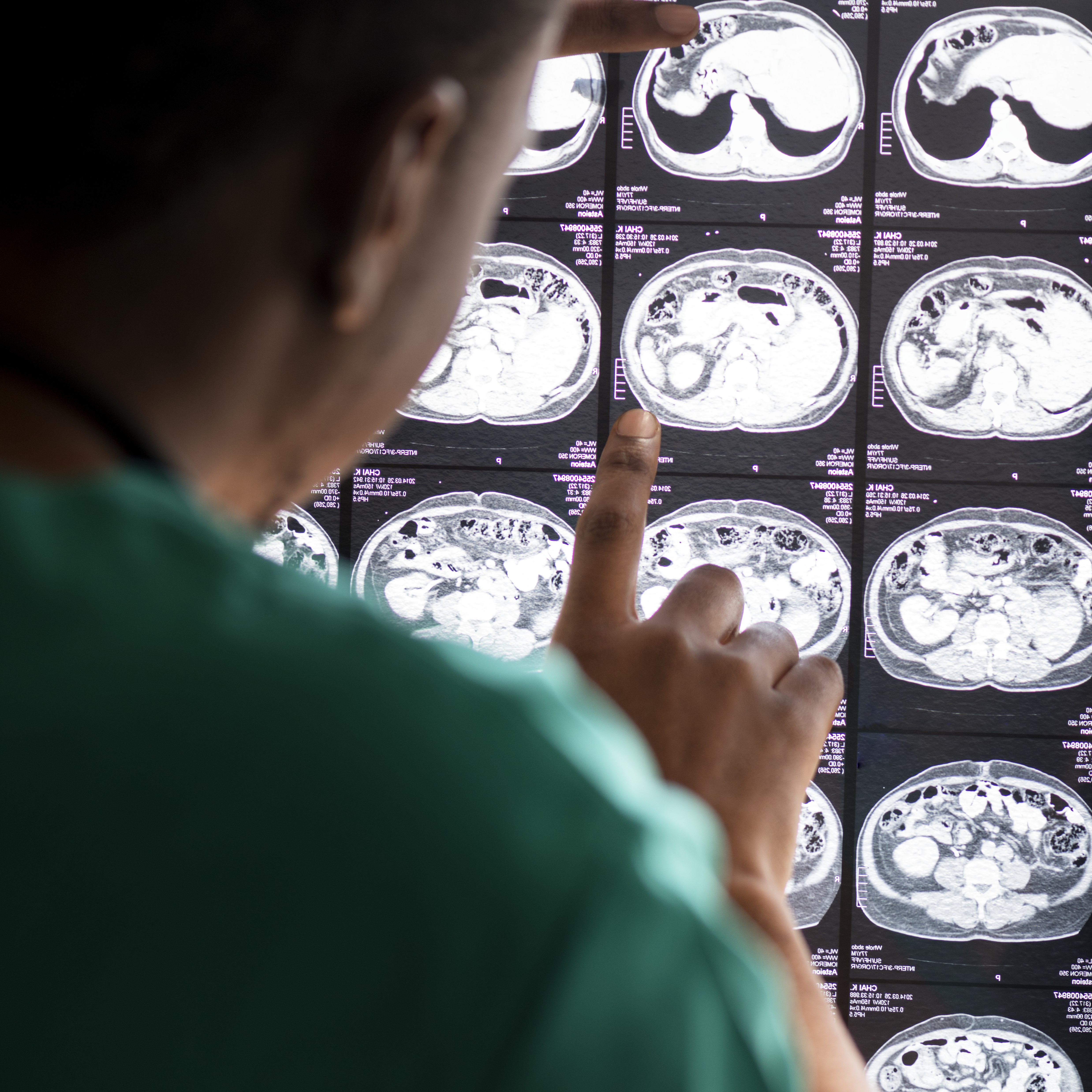
A CT scan is almost always the first test performed when a stroke is suspected. It is fast and highly effective at identifying one of the most urgent questions in stroke care: is there bleeding in the brain?
This matters enormously because the three main types of strokes:
These three types of stokes require completely different treatments. Giving clot-dissolving medication to a patient who is actually bleeding in the brain would be dangerous. A CT scan provides this critical information within minutes.
Beyond ruling out bleeding, a CT scan can also show early signs of brain tissue damage from reduced blood flow, as well as brain swelling and other structural changes.
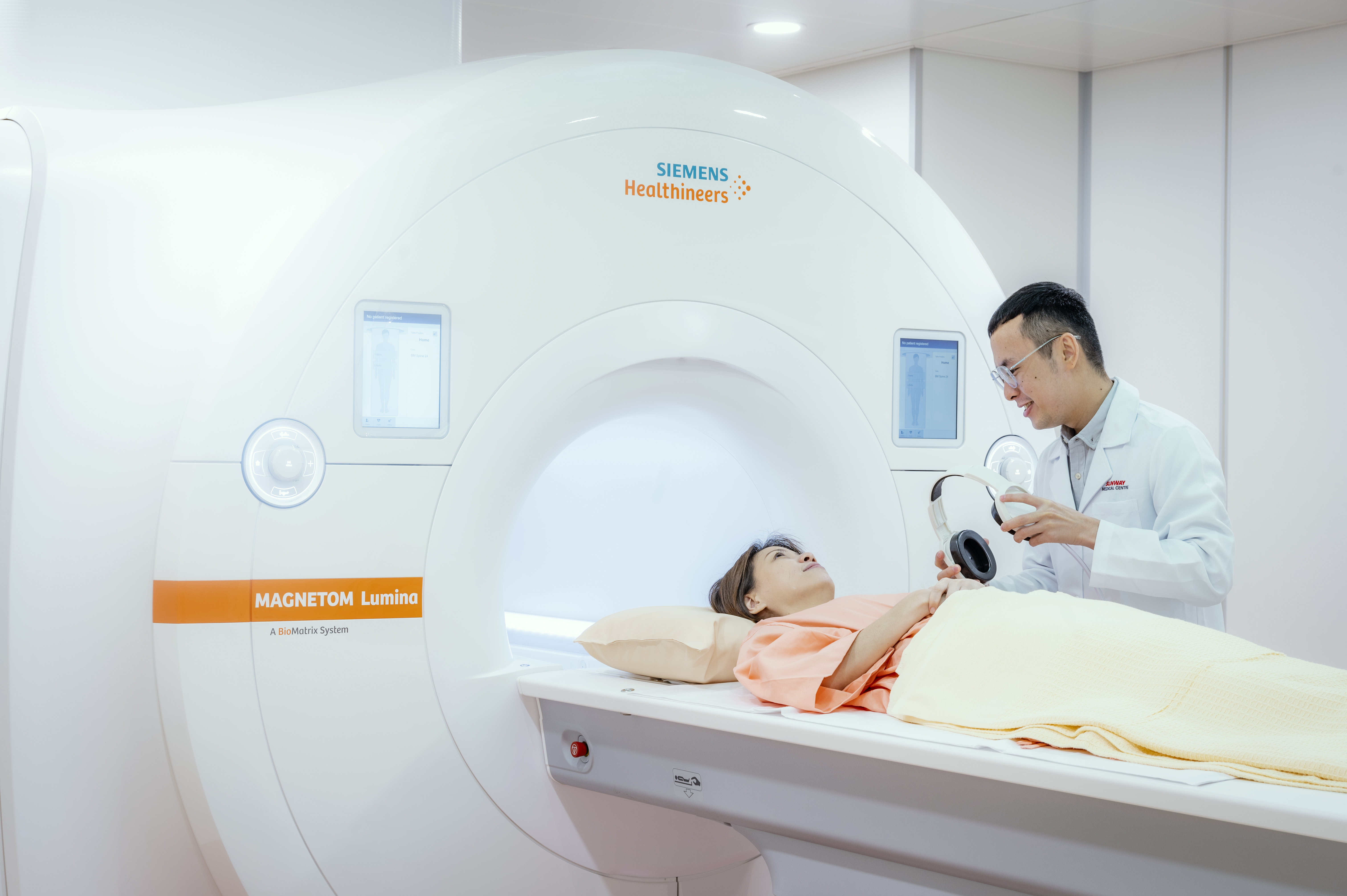
An MRI uses powerful magnetic fields and radio waves to produce highly detailed images of the brain. Unlike a CT scan, it does not use radiation. It is significantly more sensitive to early stroke damage and can detect changes in brain tissue within minutes of a stroke occurring, even when a CT scan would not yet show anything abnormal.
When used with a technique called Diffusion-Weighted Imaging (DWI), MRI is considered the gold standard for confirming acute ischaemic stroke. DWI detects the precise area of brain tissue that has been deprived of blood and oxygen, making it especially valuable for identifying small strokes deep within the brain, and strokes in the brainstem or cerebellum, which are very difficult to see on a standard CT scan.
MRA Brain is a specialised type of MRI that focuses specifically on the blood vessels in and around the brain rather than the brain tissue itself. It produces detailed images of the cerebral blood vessels without the use of radiation or contrast dye, making it a safe and non-invasive option for examining vascular structure.
MRA is used to identify narrowed, blocked, or abnormal blood vessels, including aneurysms, which are bulges in a blood vessel wall that carry a risk of rupturing, and arteriovenous malformations (AVMs), which are abnormal tangles of blood vessels that can bleed into the brain. These conditions require specific management and cannot be reliably detected on a standard CT or MRI alone.
MRA is often performed alongside MRI Brain as part of a combined neurological assessment, giving the medical team a complete picture of both the brain tissue and the blood vessels supplying it.
For patients who may need a procedure such as mechanical thrombectomy to remove a clot, cerebral angiography may also be performed. This is a more advanced imaging technique in which a thin, flexible catheter is inserted into a blood vessel in the wrist or groin and guided through to the blood vessels of the brain under live X-ray guidance. A contrast dye is then injected to produce real-time, high-resolution images of blood flow inside the brain.
At Sunway Medical Centre Penang, cerebral angiography is performed by our Consultant Interventional Radiologist inside our Hybrid Catheterisation Laboratory (Cath Lab). This is a specialised facility equipped to both diagnose and treat stroke in the same room, removing any delay between confirming the diagnosis and beginning the intervention.
Knowing that a stroke has occurred is only part of the picture. Identifying why it happened is equally important, because the cause determines what medication or intervention is needed to prevent a second stroke.
Checks the heart's electrical rhythm. This is particularly important because an irregular heartbeat called atrial fibrillation (AFib) is one of the most common causes of ischaemic stroke. When the heart beats irregularly, blood can pool and form clots that travel through the bloodstream to the brain. Identifying AFib means the right medication can be started to prevent this from happening again.
An ultrasound of the heart may also be performed. This examines the structure of the heart chambers and valves and checks for blood clots or abnormalities that may have been the source of a clot that reached the brain.
Uses sound waves to examine the carotid arteries in the neck, which are the main blood vessels carrying blood from the heart to the brain. Narrowing or plaque build-up in these arteries is a significant risk factor for stroke. Identifying this allows the team to plan treatment, whether through medication or a procedure, to reduce the risk of a future event.
You should seek emergency help immediately; the moment symptoms appear. The most effective stroke treatments have strict time windows — clot-dissolving medication must be given within 4.5 hours of symptom onset, and outcomes from clot removal procedures are significantly better the sooner they are performed. Even if symptoms seem mild or begin to improve on their own, do not wait. Call for emergency assistance right away and note the exact time the symptoms started.
At Sunway Medical Centre Penang, once a stroke is confirmed through brain imaging and clinical assessment, our stroke team moves immediately to determine the most appropriate treatment based on the type of stroke, the area of the brain affected, the severity of symptoms, and how much time has passed since onset. Every diagnostic finding directly shapes the treatment pathway that follows.
Speak with our Stroke Specialists to understand what the diagnostic process involves and what to expect next.
Learn more about stroke care at our Stroke Centre in Penang
Read More: What Is a Stroke? | Stroke Symptoms and Warning Signs | Stroke Risk Factors | Stroke Prevention | Stroke Treatment | Stroke Specialists | Stroke Rehabilitation





















Make Appointment Now
Looking for Specialist Care? Schedule a Consultation Today.